Followers

Welcome

Powered by Blogger.

About

Trying to provide all necessary information about IMMUNITY and IMMUNE SYSTEM

Immunoglobulin D(IgD),immunology,Immune system,immunity.

Posted by Mumtaz khan Wednesday, 15 February 2012 0 comments

Immunoglobulin D(IgD):
IgD was first discovered when a patient developed a multiple myeloma protein failed to react with anti-isotype antisera against the then-known isotypes:IgA,IgM,and IgG.When rabbits were immunized with this myeloma protein,the resulting antisera were used used to identify the same class of antibody at low levels in normal human serum.
The new class,called IgD,has a serum concentration of 30 mu d/ml and constitute about 0.2% of the total immunoglobulin in serum.IgD,together with IgM,is the major membrane-bound immunoglobulin expressed by mature B cells,and its role in the physiology of B cells is under investigation.No biological effector function has been identified for IgD.

| edit post

Immunoglobulin E(IgE),Immunology,Immune system,Immunity.

Posted by Mumtaz khan Tuesday, 14 February 2012 0 comments

Immunoglobulin E(IgE):
           The potent biological activity of IgE allowed it to be identified in serum despite its extremely low average serum concentration.IgE antibodies mediate the immediate hypersensitivity reactions that are responsible for symptoms of hay fever,asthma,hives,and anaphylactic shock.The presence of a serum component responsible for allergic reactions was first demonstrated in 1921 by K.Prausnitz and H.Kustner,who injected serum from an allergic person intra-dermally into a nonallergic individual.When the appropriate antigen was later injected at the same site,a wheal and flare reaction(analogous to hives)developed there.This reaction,called the P-K reaction(named for its originators,prausnitz and kustner),was the basis for the fiorst biological assay for IgE activity.

a) Cross-linkage by a single allergen and b) Cross-linkage by a single IgG antibody to two allergen molecules.
          Actual identification of IgE was accomplished  by K.prausnitz and T.Ishizaka in 1966.They obtained serum from an allergic individual and immunized rabbits with it to prepare anti-isotype antiserum.The rabbits antiserum was then allowed to react with each class of human antibody known at that time(i.e.,IgG,IgA,IgM,IgD).In this way,each of known anti-isotype antibodies was precipitated and removed from the rabbit anti-serum.What remained was an anti-isotype antibody specific for an unidentified class of antibody.This antibody turned out to completely block the P-K reaction.the new antibody was called IgE(in reference to the E antigen of ragweed pollen,which is a potent inducer of this class of antibody).
         IgE binds to Fc receptors on the membranes of blood basophils and tissue mast cells.Cross-linkage of receptor-bound IgE molecules by antigen(allergen)induces basophils and mast cells to translocate their granules to the plasma membrane and release their contents to the extracellular environment, aprocess known as degranulation.As a result,a variety of pharmacologically active mediators are released and give rise to allergic manifestations.Localized mast-cell degranulation induced by IgE also may release for antiparasitic defense.

| edit post

Immunoglobulin(IgA),Immunology,Immune system,Immunity.

Posted by Mumtaz khan Monday, 13 February 2012 0 comments


Immunoglobulin A (IgA):
            Although IgA constitutes only 10%-15% of the total immunoglobulin in serum,it is the predominant immunoglobulin class in external secretions such as breast milk,saliva,tears,and mucus of the bronchial,genitourinary,and digestive tracts.In serum,IgA exists primarily as a monomer,but polymeric forms(dimers,trimers,and some tetramers)are sometimes seen,all containing a J-chain polypeptide.The IgA of external secretions,called secretory IgA,consists of a dimer or a tetramer,a J-chain polypeptide,and a polypeptide chain called secretory component.As is explained below,secretory component is derived from the receptors that is responsible for transporting polymeric IgA across cell membranes.The J-chain polypeptide in IgA is identical to that found in pentameric IgM and serves a similar function in facilitating the polymerization of both serum IgA and secretory IgA.The secretory component is a 70,000-MW polypeptide produced by epithelial cells of mucous membranes.It also consists of five immunoglobulin-like domains that bind to the Fc region domains of the IgA dimer.This interaction is stabilized by a sulfide bond between the fifth domain of the secretory component and one of the chains of the dimeric IgA.

           The daily production of secretory IgA is greater than that of any other immunoglobulin class.IgA-secreting plasma cells are concentrated along mucous membrane surfaces.Along the jejunum of the small intestine,for example,there are so many IgA secreting plasma cells- a number that surpasses the total plasma cells population of the bone marrow,lymph and spleen combined ! Every day,a human secretes from 5 g to 15 g of secretory IgA into mucous secretions.
           The plasma cells that produce IgA preferentially migrate to sub epithelial tissue,where the secreted IgA binds tightly to receptor for polymeric immunoglobulin molecules.This poly-Ig-receptor is expressed on the basolateral surface of most mucosal epithelia(e.g.,the lining of digestive,respiratory,and genital tracts)and on glandular epithelia in the mammary,salivary,and lacrimal glands.After polymeric IgA binds to the poly-Ig receptor,the receptor-IgA complex is transported across the epithelial barrier to the lumen.Transport of the receptor-IgA complex involves receptor-mediated endocytosis into coated pits and directed transport of the vesicle across the epithelial cell to the luminal membrane,where the vesicle fuses with the plasma membrane.The poly-ig receptor is then cleaved enzymatically from the membrane and becomes the secretory component,which is bound to and released together with polymeric IgA into the mucous secretions.The secretory component masks sites susceptible to protease cleavage in the hinge region of secretory IgA,allowing the polymeric moleculs exist longer in the protease-rich mucosal environment than would be possible otherwise.Pentameric IgM is also transported into mucous secretions than does IgA.The poly-Ig receptor interact with the J- chain of both polymeric IgA and IgM antibodies.
         Secretory IgA serves an important effector function at mucous membrane surfaces,which are the main entry sites for most pathogenic organisms.Because it is polymeric,secretory IgA can cross-link large antigens with multiple epitopes.Binding of secretory IgA to bacterial and viral surface antigens prevents attachment of the pathogens to the mucosal cells,thus inhibiting viral infection and bacterial colonization.Complexes of secretory IgA and antigens are easily entrapped in mucus and then eliminated by the ciliated epithelial cells of respiratory tract or by peristalsis of the gut.Secretory IgA has been shown to provide an important line of defense against bacteria such as Salmonella,Vibrio cholerae,and Neisseria gonorrhoeae and viruses such as polio,influenza,and reovirus.
         Brast milk contains secretory IgA and many other molecules that help protect the newborn against infection during the first month of life.Because the immune system of infants is not fully functional,breast-feeding plays an important role in maintaining the health of newborns.

| edit post

Immunoglobulin M(IgM),Immunology,Immunity,Immune system

Posted by Mumtaz khan Friday, 10 February 2012 0 comments


Immunoglobulin M (IgM):      
    IgM accounts for 5%-10% of the total serum immunoglobulin,with an average serum concentration of 1.5 mg/ml.Monomeric IgM,with a molecular weight of 180,000, is expressed as membrane-bound antibody on B cells.IgM is secreted by plasma cells as a pentamer in which five monomer units are held together by disulfide bonds that link their domains.The five monomer subunits are arranged with their Fc regions in the center of the pentamer and the ten antigen-binding sites on the periphery of the molecule.Each pentamer contains an additional Fc-linked polypeptide called the J (joining)chain,which is disulfide-bonded to the carboxyl-terminal cysteine residue of two of the ten chains.The J chain appears to be required for polymerization of the monomers to form pentameric Igm;it is added just before secretion of the pentamer.
IgM is the first immunoglobulin class produced in a primary response to an antigen,and it is also the first immunoglobulin to be synthesised by the neonate.because of its pentameric structure with 10 antigen-binding sites,serum IgM has a higher valency than the other isotypes.An IgM molecule can bind 10 small hapten molecules;however,because of steric hindrance,only 5 or fewer molecules of larger antigens can be bound simultaneously.Because of its high valency,pentameric IgM is more efficient than other isotypes in binding antigens with many repeating epitopes such as viral particles and red blood cells(RBCs).For example,when RBCs are incubated with specific antibody,they clump together into large aggregates in a process called agglutination.It takes 100 to 1000 times more molecules of IgG than IgM to achievethe same level of agglutination.A similar phenomenon occurs with viral particles:less igM is required to neutralize viral infectivity.IgM is alsoefficient than IgG at activating complement.Complement activation requires two Fc regions in close proximity,and the pentameric structure of a single molecule of IgM fulfills this requirement.
         Because of its large size,IgM does not diffuse well and therefore is found in very low concentrations in the intercellular tissue fluids.The presence of J chain allows IgM  to bind to receptors on secretory cells,which transport it across epithelial linings to enter external secretions that bathe mucosal surfaces.Although IgA is the major isotype found in tese secretions,IgM plays an important accessory role as a secretory immunoglobulin.

| edit post

Antibody Classes and Biological Activities,Immunoglobulin(IgG)

Posted by Mumtaz khan Wednesday, 8 February 2012 0 comments


          The various immunoglobulin isotypes and classes have been mentioned briefly already.Each class is distinguished by unique amino acid sequences in heavy-chain constant region that confer class-specific structural and functional properties .In this section,the structure and effector functions of each class are described in more detail.

Immunoglobulin G (IgG):
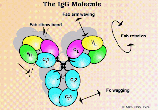
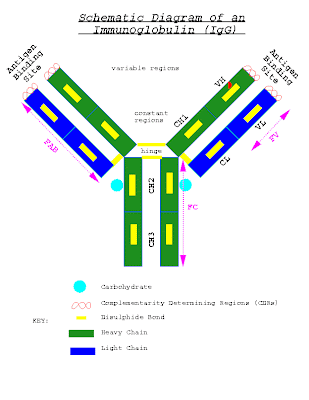
IgG,the most abundant class in serum,constitutes about 80% of the total serum immunoglobulin.The IgG molecule consists of two heavy chains and two light chains.There are four human IgG subclasses,distinguished by differences in heavy-chain sequence and numbered according to their decreasing average serum concentrations:IgG1,IgG2,IgG3,and IgG4.

The amino acid sequence that distinguish the four IgG subclasses are encoded by different germ-line genes,whose DNA sequences are 90%-95% homologous.The structural characteristics that distinguish these subclasses from one another are the size of the hinge region and the number and position of the interchain disulfide bonds between the heavy chains.The subtle amino acid differences between subclasses of IgG affect the biological activity of the molecule:
* IgG1,IgG3,and IgG4 readily cross the placenta and play an important role in protecting the developing fetus.
* IgG3 is the most effective complement at all.
* IgG1 and IgG3 bind with high affinity to Fc receptors on phagocytic cells and thus mediate opsonization.IgG4 has an intermediate affinity for Fc receptors,and IgG2 has an extremely low affinity.

| edit post

Immunoglobulin Fine structure|Immunology,Immune system,Immunity

Posted by Mumtaz khan Tuesday, 7 February 2012 0 comments

           The structure of the immunoglobulin molecule is determined by the primary,secondary,tertiary, and quaternary organization of the protein.
The primary structure,the amino acid sequence,accounts for the variable and constant regions of the heavy and light chains.The secondary structure is formed by folding of the extended polypeptide chain back and forth upon itself into an antiparallel beta pleated sheet.

The chains are then folded into tertiary structure of compact globular domains by continuations of the polypeptide chain that lie outside the beta pleated.Finally,the globular domains of adjacent heavy and light polypeptide chains interact in the quaternary structure,forming functional domains that enable the molecule to specifically bind antigen and,at the same time,perform a number of biological effector functions.

| edit post

Hematopoiesis|Formation of blood cells|Immune system.

Posted by Mumtaz khan Monday, 6 February 2012 0 comments


Hematopoiesis

        All blood cells arise from a type of cell called the hematopoietic stem cell(HSC).Stem cells are cells that can differentiate into other cell type;they are self renewing-thy maintain their population level by cell division.In humans,hematopoeisis,the formation and development of red and white blood cells,begins in the embryonic yolk sac during the first weeks of development.Here,yolk sac stem cells differentiate into primitive erythroid cells that contain embryonic hemoglobin.In the third month of gestation, hematopoietic stem cells migrate from the yolk sac to the fetal liver and then to the spleen;these two organs have major roles in hematopoiesis from third to the seventh months of gestation.After that,the differentiation of HSCs in the bone marrow becomes the major factor in hematopoiesis,and by birth there is little or no hematopoiesis in the liver and spleen.
       It is remarkable that every functionally specialized,mature blood cell is derived from the same type of stem cell.In contrast to a uni-potent cell,which differentiates into a single cell type,a hematopoeitic stem cell is multipotent,or pluripotent,able to differentiate in various ways and thereby generate erythrocytes,granulocytes,monocytes,mast cells,lymphocytes,and megakaryocytes.These stem cells are few,normally fewer than one HSC per 5 X 100000 cells in the bone marrow.
      The study of hematopoietic stem cells is difficult bot because of their scarcity and because they are hard to grow in vitro.As a result,little is known about how their proliferation for self-renewal,hematopoietic stem cells are maintained at stable levels throughout adult life;however,when there is an increased demand for hematopoiesis,HSCs display an enormous proliferative capacity.This can be demonstrated in mice whose hematopoietic systems have ben completely destroyed by a lethal dose of x-rays.Such irradiated mice will die within 10 days unless they are infused with normal bone-marrow cells from a syngeneic(genetically identical) mouse.
        Early in hematopoiesis, a multipotent stem cell differentiates along one of two pathways,giving rise to either a common lymphoid progenitor cell or common myeloid progenitor cell.The types and amounts growth factors in the microenvironment of a particular stem cell or progenitor cell control its differentiation.During the development of the lymphoid and myeloid lineages,stem cells differentiate into progenitors cells,which have lost the capacity for self-renewal and are committed to a particular cell lineage.Common lymphoid progenitor cells give rise to B,T,and NK(natural killer)cells and some dendritic cells.Myeloid stem cells generate progenitors of red blood cells(erythrocytes),many of the various white blood cells(neutophils,eosinophils,basophils,monocytes,mast cells,dendritic cells.),platelets.Progenitors commitment depends on the acquisition of responsiveness to particular growth factors and cytokines.when the appropriate factors and cytokines are present,progenitor cells proliferate and differentiate into corresponding cell type,either a mature erythrocyte,a particular type of leukocyte,or a platelet-generating cell(the megakaryocyte).Red and white blood cells pass into bone-marrow channels,from which they enter the circulation.
        In bone marrow,hematopoietic cells grow and mature on a meshwork of stromal cells,endothelial cells,fibroblasts,and macrophages.Stromal cells influence the differentiation of hematopoietic stem cells by providing a hematopoietic-including micro-environment (HIM) consisting of a cellular matrix and factors are soluble agents that arrive at their target cells by diffusion,others are membrane-bound molecules on the surface of stromal cells that require cell-to-cell contact between the responding cells and the stromal cells.During infection,hematopoiesis is stimulated by the production of hematopoietic growth factors by activated macrophages and T cells.

| edit post