Followers

Welcome

Powered by Blogger.

About

Trying to provide all necessary information about IMMUNITY and IMMUNE SYSTEM

Lymphoid Cells and Organs- Evolutionary comparisons

Posted by Mumtaz khan Friday, 30 December 2011 0 comments

         While innate systems of immunity are seen in invertebrates and even in plants,the evolution of lymphoid cells and organs evolved only in the phylum Vertebrata.Consequently,adaptive immunity,which is mediated by antibodies and T cells,is only seen in this phylum.


As one considers the spectrum from the earliest vertebrates,the jawless fishes(agnatha),to the birds and mammals,evolution has added organs and tissues with immune functions but has tended to retain those evolved by earlier orders.While all have gut-associated tissue (GALT)and most have some version of a spleen and thymus,not all have the ability to form germi centers is not shared by all.The differences seen at the level of organs and the tissues are also reflected at the cellular level.Lymphocytes that express antigen specific receptors on their surfaces are necessary to mount an adaptive immune response in lampreys and hagfish,members of order Agnatha,have failed.In fact,only jawed vertebrates ,of which the cartilaginous fish(sharks,rays)are the earliest example,have b and T lymphocytes and support adaptive immune responses.

| edit post

Flow cytometry and Leukemic typing

Posted by Mumtaz khan Thursday, 8 December 2011 1 comments

LEUKEMIA is the unchecked proliferation of an abnormal clone of hematopoietic cells.Typically,leukemic cells respond poorly or inappropriately to regulatory signals,display aberrant patterns of differentiation,or even fail to differentiate.Furthermore,they sometimes supress the growth of normal lymphoid and myeloid cells.Leukemia can rise at any maturational stage of any one of the hematopoeitic lineages.Lymphocytic leukemias display many characteristics of cells of the lymphoid lineage;another broad group,myelogenous leukemias have attributes of members of the myeloid lineage.Aside from lineage,many leukemias can be classified as cute aor chronic.Some ezamples are acute lymphocytic leukemia(ALL),the most common childhood leukemia;acute myelogenous leukemia;acute myelogenous leukemia(AML),found more often in adults than in children;and chronic lymphocytic leukemia(CLL),which is rarely seen in children but is the most common form of adult leukemia in the Western world.A fourth type,chronic(CML),occurs much more often in older adults than in children.

The diagnosis of leukemia is made on the basis of two findings.One is the detection of abnormal cells in the blood-stream,and other is observation of abnormal cells in the bone marrow.Clinical experience has shown that designing the most appropriate therapy for the patient requires knowing which type of leukemia is present.In this regard,two of the important questions are: (1)What is the lineage of the abnormal cells and (2)What is their maturational stage? A variety of approaches,including cytologic examination of cell morphology and staining characteristics,immuno-phenotyping,and,in some cases,an analusis of gene rearrangements,are useful in answering these questions.One of the most powerful of these approaches is immunophenotyping,the determination of the profile of selected cell-surface markers displayed by the leukemic cell.Although leukemia-specific antigens have not yet been found,profiles of expressed surface antigens often frequently helpful in determining the maturational stages present in leukemic cell populations.For example,an abnormal cell that displays surface immunoglobin would be assigned to the B-cell lineage and its maturational stage would be that of a mature B cell.On the other hand,a cell that had cytoplasmic heavy chains,but no surface immuno-globin,would be a B-lineage leukemic cell but at the maturational stage of a pre-B cell. The most efficient and prcise technology for immunophenotyping uses flowcytometry and monoclonal antibodies.The availability of antibodies specific for each of the scores of antigens found on various types and subtypes of hematopoietic cells has made it possible to identify patterns of antigen expression that are typical of cell lineages,maturational stages,and a number of different types of leukemia.Most cancer centres are equipped with flow cytometers that are capable of performing and interpreting the multiparameter analyses necessary to provide useful profiles surface markers on tumor cell populations.Flow cytometric determination of immuno-phenotypes allows:
* Confirmation of diagnosis
* Diagnosis when no clear judgement can be made based on morphology or patterns of cytochemical staining.
* Identification of aberrant antigen profiles that can help identify the return of leukemia during remission
* Improved prediction of the course of the disease.

| edit post

Cutaneous-Associated Lymphoid Tissue..

Posted by Mumtaz khan Tuesday, 29 November 2011 2 comments


The skin is an important anatomic barrier to the external environment,and its large surface area makes this tissue important in nonspecific(innate)defenses.The epidermal (outer)layer of the skin is composed largely of specialized epithelial cells called keratinocytes.These cells secrete a number of cytokines that may function to induce a local inflammatory reaction.In addition,keratinocytes can be induced to express class II MHC molecules and may function as antigen-presenting cells.Scattered among the epithelial-cell-matrix of the epidermis are Langerhans cells,a type of dendritic cell,which internalize antigen by phagocytosis or endocytosis.The Langerhans cells then migrate from the epidermis to regional lymph nodes,where they differentiate into interdigitating dendritic cells.These cells express high levels of class II MHC molecules and function as potent activators of naive Helper cells.
 
The epidermis also contains so-called intraepidermal lymphocytes.These are similar ti the intraepithelial lymphocytes of MALT in that most of them are CD8 plus cells,which have limited diversity for antigens that enters through the skin and some immunologist believe that they may play a role in combating antigens that enter through skin.The under-laying dermal layer of the skin contains scattered CD4 plus and CD8 plus T cells and macrophages.Most of these dermal T cells were either previously activated cells or are memory cells.

| edit post

Mucosa-Associated Lymphoid Tissue.

Posted by Mumtaz khan Monday, 21 November 2011 2 comments

 MUCOSA-ASSOCIATED LYMPHOID TISSUE

The mucous membranes lining the digestive,respiratory,and urogenital systems have a combined surface area of about 400 metre per square(nearly the size of a basket ball court)and are the major sites of entry for most pathogens.These vulnerable membrane surfaces are defended by a group of organized lymphoid tissues mentioned earlier and known collectively as mucosa-associated lymphoid tissue (MALT). Structurally,these range from loose,barely organized clusters of lymphoid cells in the lamina propria of intestinal villi to well-organized structures such as the familiar tonsils and appendix,as well as Peyer's patches,which are found within the submucosal layer of the intestinal lining.The functional importance of MALT in the body's defense is attested to by its large population of antibody-producing plasma cells,whose number far exceeds that of plasma cells in the spleen,lymph nodes,and bone marrow combined.

The tonsils are found in three locations:lingual at the base of the tongue;palatine at the sides of the back of the mouth;and pharyngeal(adenoids) in the the roof of the nasopharynx.All three tonsil groups are nodular structures consisting of a meshwork of reticular cells and fibers interspersed with lymphocytes,macrophages,granulocytes and mast cells.The B cells are organized into follicles and germinal centers; the latter are surrounded by regions showing T-cell activity.The tonsils defend against antigens entering through the nasal and oral epithelial routes.
 The best studied of the mucous membranes is the one that lines the gastrointestinal tract.This tissue,like that respiratory and urogenital tracts,has the capacity to endocytose antigen from the lumen.Immune reactions are initiated against pathogens and antibody can be generated and exported to the lumen to combat the invading organisms.The lymphoid cells are found in various regions within this tissue.The outer mucoal epithelial layer contains so-called intraepithelial lymphocytes(IELs).Many of these lymphocytes ate T cells that express unusual receptors,which exhibit limited diversity for antigen.Although this population of T cells is well situated to encounter antigens that encounter through the intestinal mucous epithelium.their actual function remains largely unknown.The lamina propria,which lies under the epithelial layer,contains large numbers of B cells,plasma cells,activated T-Helper cells,and macrophages in loose clusters.Histologic sections have revealed more than 15000 lymphoid follicles within the intestinal lamina propria of a healthy child.The submucosal layer beneath the lamina propria contains Peyer's patches,nodules of 30-40 lymphoid follicles in other sites,those that compose Peyer's patches can develop into secondary follicles with germinal centers.
The epithelial cells of mucous membranes play an important role in promoting the immune response by delivering small samples of foriegn antigen from the lumina of the respiratory,digestive,and urogenital tracts to the underlaying mucosal-associated lymphoid tissue.This antigen transport is carried out by specialized M cells.

Structure of M cells :
Structure of M cells and production of IgA at inductive sites.
         The structure of M cells is striking: These are flattened epithelial cells lacking the microvilli that characterize the rest of the mucous epithelium.In addition,M cells have deep invagination,or pocket,in the basolateral plasma membrane;this pocket is filled with cluster of B cells,T cells,and macrophages.Luminal antigens are endocytosed into vesicles that are transported from the luminal membrane to the underlying pocket membrane .The vesicle then fuse with the pocket membrane,delivering the potentially response-activating antigens to the clusters of lymphocytes contained within the pocket.
         M cells are located in so-called inductive sites-small regions of a mucous membrane that lie over organized lymphoid follicles.Antigens transported across the mucous membrane by M cells can activate B cells within these lymphoid follicles.The activated B cells differentiate into plasma cells,which leave the follicles and secrete the IgA into the lumen,where they can interact with antigens.            
        Mucous membranes are an effective barrier to the entrance of most pathogens,which thereby contributes to nonspecific immunity.One reason for this is that the mucosal epithelial cells are cemented to one another by tight junctions that make it difficult for pathogens,including both bacteria and viruses,have exploited the M cell as an entry route through the mucous-membrane barrier.In some cases,the pathogen is internalized by the M cell and transported into the pocket.In other cases,thus allowing entry of the pathogen.Among the pathogens that use M cells in these ways are several invasive Salmonella species,Vibrio cholerae,and the polio virus.

| edit post

Spleen-Role of Spleen in immune system

Posted by Mumtaz khan Friday, 18 November 2011 0 comments

SPLEEN:
     The spleen plays a major role in mounting immune response to antigens in the blood stream.It is a large,ovoid secondary organ sitauted high in the left abdominal cavity.While lymph nodes are specialized for trapping antigen from local tissues,the spleen specializes in filtering blood and trapping blood-borne antigens;thus ,it can reepond to systemic infections.Unlike the lymph nodes,the spleen is not supplied by lymphatic vessels.Instead,blood-borne antigens and lymphocytes are carried into spleen through the splenic artery.

       The spleen is surrounded by a capsule that extends a number of projections(trabaculae)into the interior to form a compartmentalized structure.The compartments are of two types,the red pulp and white pulp,which are separated by a diffuse marginal zone.The splenic red pulp consists of a network of sinusoids populated by macrophages and numerous red blood cells(erythrocytes)and few lymphocytes;it is the site where old and defective red blood cells are destroyed and removed.Many of the macrophages within the red pulp contain engulfed red blood cells or iron pigments from degraded hemoglobin.The splenic white pulp surrounds the branches of the splenic artery,forming a periarteriolar lymphoid sheath(PALS)populated mainly by T lymphocytes.Primary lymphoid follicles are attached to thePALS.These follicles are rich in B cells and some of them contain germinal centers.The marginal zone,located peripheral to the PALS,is populated by lymphocytes and macrophages.

| edit post

Lymph nodes-Role in Immune response

Posted by Mumtaz khan Thursday, 17 November 2011 0 comments

LYMPH NODES: 

         Lymph nodes are the sites where immune responses are mounted to antigens in lymph.They are encapsulated bean-shaped structures containing a reticular packed with lymphocytes,macrophages,and dendritic cells.Clustered at junctions of the lymphatic vessels,lymph nodes are the first organized lymphoid structure to encounter antigens that enter the tissue spaces.

Morphologically, node can be divided into three roughly concentric regions:the cortex,the paracortex and the medulla,each of which supports a distinct microenvironment. The outermost layer,the cortex,contains lymphocytes(mostly B cells),macropahges,and follicular dendritic cells arranged in primary follicles.After antigenic challenge,the primary follicles enlarge into secondary follicles ,each containing a germinal center.In children with B-cell deficiencies,the cortex lacks primary follicles and germinal centers.Beneath the cortex is the paracortex,which is populated largely by T lymphocytes and also contains interdigitating dendritic cells thought to have migrated from tissues to the node.These interdigitating dendritic cells express high levels of class II MHC molecules,which are necessary for presenting antigen. Lymph nodes taken from neonatally thymectomized mice have unusually few cells in the paracortical region; the paracortex is therefore sometimes referred to as thymus-independent area in contrast to the cortex,which is a thymus-independent area.The innermost layer of a lymph node,the medulla,is more sparsely populated with lymphoid-lineage cells;of those present,many are plasma cells actively secreting antibody molecules.

FUNCTIONS OF LYMPH NODES :
         This work suggests an explanation for the curious fact that patients receiving a liver transplant sometimes inherit the donor’s allergies and immune repertoire, so in keeping with the idea that donor immune information is being transplanted. It also suggests that the liver as an immune organ is an evolutionary remnant from the time before lymph nodes developed in higher birds and mammals. Cold-blooded vertebrates have functioning T and B cells but no lymph nodes. The main achievement of the development of lymph nodes in mammals is a drastic improvement for the production of better antibodies. T cells on the other hand have not changed their function much during evolution and the work by the Zurich group finally provides solid evidence for the versatility and promiscuity of this cell type.


| edit post

Bone marrow-Role played in Immune ayatem

Posted by Mumtaz khan Wednesday, 16 November 2011 0 comments

             BONE MARROW
              In humans and mice,bone marrow is the site of B-cell origin and development.Arising from lymphoid progenitors,immature B cells proliferate and differentiate within the bone marrow, and stromal cells within interact directly with B cells and secrete various cytokines that are required for development.


Bone marrow is not  the site of B-cell development in all species..In birds,a lymphoid organ called the bursa of fabricus,a lymphoid tissue associated with the gut,is the primary site of B-cell maturation,proliferation.In mammals such as primates and rodents,there is no bursa and no single counterpart to it as a primary lymphoid organ.In cattle and sheep,the primary lymphoid tissue hosting the maturation the maturation,proliferation,and diversification of B cells early in gestation is the fetal spleen.


Later in gestation,this function is assumed by a patch of tissue embedded in the wall of the intestine called the ileal Peyer's patch,which contains a large number of B cells.The  rabbit,too,uses gut-associated tissue suchas the appendix as primary lymphoid tissue for important steps in the proliferation and diversification of B cells.


FUNCTION OF BONE MARROW:
The bone marrow is found within the central cavities of axial and long bones. It consists of hematopoietic tissue islands and adipose cells surrounded by vascular sinuses interspersed within a meshwork of trabecular bone. It accounts for approximately 3% of the body weight in adult rats , ~2% in dogs and ~5% in humans . The bone marrow is the major hematopoietic organ, and a primary lymphoid tissue, responsible for the production of erythrocytes, granulocytes, monocytes, lymphocytes and platelets.

| edit post


 THYMUS
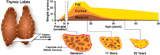
               The Thymus is the site of T-cell and maturation.It is a flat,bilobed organ situated above the heart.Each lobe is surrounded by a capsule and is divided into lobules,which are separated from each other by strands of connective tissue called trabaculae.Each lobule is organized into two compartment,or cortex,is densely packed with immature T cells,called thymocytes,whereas the inner compartment,or medulla,is sparsely populated with thymocytes.
Diagrammatic cross section of portion of thymus.
               Both the cortex and medulla of the thymus are criss-crossed by a three-dimensional stromal-cell network composed of epithelial cells,dendritic cells, and macrophages,which make up the framework of the organ and contribute to the growth and maturation of the thymocytes.Many of these stromal cells interact physically with the developing thymocytes.Some thymic epithelial cells in the outer cortex,called nurse cells,have long membrane extension that surround as many as 50 thymocytes,forming large multicellular complexes.Other cortical epithelial cells have long interconnecting cytoplasmic extensions that form a network and have been shown to intreract with numerous thymocyets  as they traverse the cortex.
               The function of the thymus is to generate and select a repertoire of T cells that will protect the body from infection.As thymocytes develop,an enormous diversity of T cell recptors is generated by a random process that produce some T cells with receptors capable  of recognizing antigen-MHC complexes.However,most of the T-cell-receptors produced by this random process are incapable recognizing antigen-MHC complexes and a small portion react with combinations of self antigen-MHC complexes.The thymus induces the death ot those that react with self-antigen-MHC and pose a danger of causing autoimmune disease.

THE THYMUS AND IMMUNE FUNCTION:

Changes in Thymus with age.
            Aging is accompanied by a decline in thymic function.This decline may play some role in the decline in immune function during aging in humans and mice.The thymus reaches its maximal size at puberty and then atrophies,with a significant decrease in both cortical and medullary cells and an increase in the total fat content of the organ.Whereas the average weight of the thymus is 70 g in infants.its age dependent involution leaves an organ with an average weight of only 3 g in the elderly.

| edit post

Organs of the Immune system

Posted by Mumtaz khan Tuesday, 15 November 2011 0 comments

Organs of the Immune system:
           A number of morphologically and functionally diverse organs and tissues have various functions in the develpoment of immune responses.These can be distinguished by functions as the primary and secondary lymphoid organs.The thymus and bone marrow are the primary(or central) lymphoid organs,where maturation of lymphocytes takes place.The lymph nodes,spleen,and various mucosal-associated lymphoid tissue(MALT) such as gut-associated lymphoid tissue(GALT) are the secondary (or peripheral)lymphoid organs,which trap antigen and provide sites for mature lymphocytes to interact with that antigen.In addition,tertiary lymphoid tissues,which normally contain fewer lymphoid cells than secondary lymphoid organs,can import lymphoid cells during inflammatory response.Most prominent of these are cutaneous-associated lymphoid tissues.Once mature lymphocytes have been generated in the primary lymphoid organs,they circulate in the blood and lymphatic system,a network of vessels that collect fluid that has escaped into the tissues from capillaries of the circulatory system and ultimately return it to blood.

| edit post

Anemia-a blood disorder

Posted by Mumtaz khan 3 comments

ANAEMIA

            Anemia is a condition in which Oxygen carrying capacity of blood is reduced.It should be noted that it is a sign and not the diagnosis.Many kinds of anemia exist.All of them are characterized by the reduced number of R.B.Cs or decreased amount of hemoglobin in the blood.These conditions lead to fatigue and intolerance to cold both of which are related to lack of oxygen needed for ATP and heat production and to paleness which is due to the low hemoglobin content.
TYPES OF ANEMIA:
Nutritional Anemia: It arises from inadequate diet usually a diet without enough Iron or a diet without vitamin B12.
Pernicious Anemia: It is the insufficient hematopoeisis that results from inability ot the stomac mucosa to produce untrinsic factor which is needed for the absorption of vitamin B12.
Haemorrhagic Anemia:An execessive loss of R.B.Cs through bleeding is called Haemorrhagic anemia.The common causes are large wounds,stomach ulcer and heavy menstrual bleeding.If the bleeding is extraordinarily heavy the anemia is turned as ''acute''.The excessive blood loss can be fatal and if bleeding is slow and prolonged then the anemia is turned as''chronic''.One of the chief symptoms is fatigue.
Haemolytic Anemia:If the cell membrane of R.B.Cs ruptures prematurely the R.B.Cs remain as ghost and their hemoglobin pores out into plasma.A characteristic sign  of this condition called hemolytic anemia is a distortion in shape of R.B.Cs.It may result from inherent defects such as a defect in the structure of hemoglobin or a defect in the cell membrane of R.B.Cs.The agents that may cause such type of anemia are parasites,toxins and antibodies coming from incompatible blood haemolytic disease of new born i.e.,erythroblastosis feotalis is an excellent example of hemolytic anemia.
Aplastic Anemia:Destruction or exhibition of red bone marrow results in Aplastic Anemia typically the bone marrow is replaced by fatty tissue or fibrous tissue or tumor cells.Medication that inhibit  or prevent the enzymes involved in hematopoeisis or radiation or certain toxins may be the causes for such type of anemia.

| edit post

Fibrinolysis and Anemia

Posted by Mumtaz khan Saturday, 12 November 2011 0 comments

FIBRINOLYSIS


               The fibrinolytic system provides ,checks and balances so that clotting does not get out of hand.It also dissolves the clot once the damage is repaired.Dissolution of clot is called Fibrinolysis.When a clot is finally formed inactive enzyme in the plasma called plasminogen gets incorporated into the blood clot.Activators from the blood and tissue now get released to activate the inactive enzyme plasminogen into the active enzyme called plasmin.Once plasma is formed,it can dissolve the blood clot by digesting the fibrin threads.

| edit post

COAGULATION FACTOR

Posted by Mumtaz khan Thursday, 10 November 2011 0 comments

COAGULATION FACTOR (c.f.):


  1. c.f I- Fibrinogen
  2. c.f  II- Prothrombin
  3. c.f III - Tissue factor /Thromboplastin
  4. c.f - IV - Calcium ions 
  5. c.f V - Proaccelerin (labile factor)
  6. c.f VII - Serum Prothrombinconversion accelarator.
  7. c.f VIII - Anti haemophilic factor (AHF)
  8. c.f  IX- Christmas factor,plasma thromboplastin component (PTC) stuart factor, Thrombokinase
  9. c.f XI - Plasma Thromboplastin antecedent (PTA)
  10. c.f  XII -Hageman factor,contact factor
  11. c.f  XII - Fibrin stabilizing factor,Fibrinase.

NOTE: There is no factor VI. Prothrobinase is a combination of activated factors V and X .

| edit post

INTRINSIC PATHWAY:
 
         The intrinsic pathway of blood clotting is more complex than extrinsic and it occurs more slowly. It is so named because its activators are in direct contact with blood.In other words,the activators are in direct contact with blood.In other words the activators are within or intrinsic to blood.If the endothelial cells become damaged,blood can come in contact with the exposed collagen.In addition the trauma to the endothelial cells causes damage to the blood platelets resulting in the release of phospholipids by the platelets contact with collagen activates c.f  XII.Activated c.f XII further activates c.f.XII Activation of c.f XI in presence of co-factor Calcium ions activates c.f IX.activation of c.f IX under the influence of co-factor Calcium ions,c.f VIII and phospholipids of platelets will activate c.f X. Activation of c.f X in combination with c.f V again under the influence of co-factor Calcium results in formation of Prothombinase.This takes several minutes.As a result completion of stage I in intrinsic pathway is of longer duration as compared to that in Extrinsic pathway.
Stage 2 and stage 3 are similar as described for Extrinsic pathway.
The last step involved which is common to both the pathways is conversion of loose fibrin threads into more stabilizing and strengthened fibrin threads is done by activated c.f XIII. C.f. XIII is activated by thrombin,then at the end the clot becomes sturdy whereby blood corpuscles have been trapped in the meshwork of stabilized fibrin in threads and what oozes out is watery straw coloured fluid serum which is plasma protein and other coagulation factor.

CLOT RETRACTION:
Once a clot is formed it plugs the ruptured area of blood vessel and prevents bleeding hemorrhage. Clot retraction or syneresis is consolidation or tightening of the fibrin clot. The fibrin threads attached to the damaged surface of blood vessel gradually contract.As the clot retracts it pulls the edges of damaged blood vessels closer and thus the risk of hemorrhage is further decreased and during this retraction some serum escapes betweeen the fibrin threads.
At times clotting mechanism starts at site which may spread beyond the area of damaged blood vessels.This is because thrombin has 2 positive feedback effects,whereby it may directly activate c.f V or result in release of phospholipids from the activated platelets from activated platelets.The combined effect of these resulting in synthesis of prothrombinase which in turn accelarates more and more synthesis of thrombin.If this remains unchecked a clot would continue to get larger and larger.Normally,however fibrin has the ability to inactivate much of the thrombin formed and this helps to stop the spread of thrombin into blood :Thus limiting the spread of the blood clot beyond the site of damage.
 

| edit post

Blood clotting mechanism

Posted by Mumtaz khan Friday, 21 October 2011 0 comments

HOW DOES BLOOD CLOT ?  
  
        Clotting is a much complex process in which all the coagulation factor must synchronize and activate each other.Basically the entire mechanism of clotting can be explained in 3 steps.
1. Formation of Prothombinase .
2. Conversion of plasma protein-prothombin into an enzyme called Thrombin by the activator-Prothombinase.
3. Conversion of another plasma protein called Fibrinogen which is soluble,into insoluble Fibrin under the influence of Thrombin.

EXTRINSIC PATHWAY:-
The extrinsic pathway of blood clotting has few steps as compared to the intrinsic pathway .This is because of clotting comes into play.When there is a severe trauma and occurs rapidly.It has been named Extrinsic because a tissue factor which is a tissue protein i.e.,coagulation factor III popularly referred as Thromboplastin leaks into the blood from the tissue cells which are outside the blood vessels(so extrinsic).This coagulation factor III is a complex mixture of Lipoprotein and phospholipid which gets released from the surface of damaged cells.Since the factor is released from the site of injury itself the 3 stage process of clotting will be rapid.This Thromboplastin activates coagulation factor VII in presence of Calcium ions.Coagulation factor VII gets activated w+hich again in the presence of Calcium ions activates CF X.Once CF X is activated it combines w+ithCF V again in the presence of Calcium ions form active enzyme Prothombinase.Formation of Prothombinase satisfies the 1st stage process in blood clotting.
Stage 2 and stage 3 processes i.e.,conversion of plasma protein Prothrombin into enzyme Thrombin by using prothrombinase and finally conversion of soluble plasma proteins fibrinogen into insoluble threads fibrin by using thrombin are the next steps completing the Extrinsic pathway.

| edit post

Coagulation of blood

Posted by Mumtaz khan Monday, 17 October 2011 0 comments

Homeostasis:-
Homeostasis refers to stoppage of bleeding.when blood vessels are ruptured or damaged hemostatic response must be quick localised to the region of damage and carefully controlled three basic mechanisms present blood loss viz,
(1) Vascular spasm.
When the blood vessels are damaged the circularly arranged smooth muscles immediately contracts this is referred to as vascular spasm and will reduce blood loss.This is crucial because during this time other homeostatic mechanism get into operation.The vascular spasm is caused by damage to the smooth muscles and reflexes initiated by pain receptors.


(2) Platelet plug formation.
The blood platelets in their original unstimulated state are disc shaped showing two types of granules in their cytoplasm viz., alpha granules and dense granules.
In the first phase of platelet formation,blood platelet come together make a contact and stick to the part of damaged blood vessels.This process is called platelet adhesion.As a result of adhesion,the platelets which were earlier unstimulated now become activated and their charactreistics change drastically.They extend projections which help them to make further contacts with the neighboring platelets and to release  their contents from the granules.This is referred to as platelet release reaction.The liberated ATP will activate the neighboring platelets as well. Serotonin  alongwith a factor called thromboxane causes vasoconstriction.Thus decreasing the bloodflow..The release of ADP will make the platelets more sticky and this stickiness will cause them to adhere to each other. This gathering of platelets for stickiness is referred to as Platelet Aggregation.

(3) Blood clotting.
 Normally blood remains in th liquid state as long as it stays within the blood vessels but if it is withdrawn from the body it gets converted from the fluid state into a gel state.Eventually the gel separates from the liquid and a yellow colored or straw colored liquid which oozes out from blood clot,is called Serum(Serum means plasma clotting proteins). The remaining gel is called a clot consisting of network of insoluble fibers of fibrin in which blood corpuscles get trapped.This process of gel formation is called coagulation or clotting.
    Clotting involves several enzymes and other chemicals known as Clotting factors.Most of these clotting factors are synthesized in the liver and finally released in the blood plasma.There are some clotting factors which is released by blood platelets and some clotting factor (recently accepted)which is released from the damaged tissue cells earlier it was called as Thromboplastin.


| edit post

HAEMATOLOGY

Posted by Mumtaz khan Monday, 10 October 2011 0 comments

HEMOGLOBIN STRUCTURE
         Hemoglobin is the major oxygen carrying molecule was the first oligomeric protein to be studied by X- ray diffraction methods.

         Two major types of hemoglobin occur in humans at different stages of development namely,Feotal Hemoglobin(Hb F),and Adult hemoglobin(Hb A). Other forms of hemoglobin such as embryonic and minor adult forms do exist.
        Each molecule of hemoglobin is composed of four polypeptide chains plus a heam group.hemoglobin A contains two identical alpha chains and two identical beta-chains.There is another type of hemoglobin A which contains sigma chains.
        Before birth several additional hemoglobin polypeptides are synthesized e.g., In early embryonic life hemoglobin show presence of Epsilon chain while in the fetal life it shows the presence of two alpha chains.While in the fetal life it shows the presence of two alpha chains and two sigma chains because both hemoglobin in embryonic life and hemoglobin in fetal life have grater affinity for oxygen than hemoglobin in adult life.The feotus can prefentially absorb from maternal blood streams.
       Each peptide chain whether alpha,beta or sigma is encoded by a specific gene.Hb F present in developing feotus is normally replaced by Hb Awithin the 1st 6 monthsafter birth.Each polypeptide consists of specific sequence of a specific sequence of AA's.

| edit post

Drug Allergies - When Medicines become Immunogens

Posted by Mumtaz khan Thursday, 29 September 2011 0 comments

     Since World War II,penicillin has been used to successfully treat a wide variety of bacterial infections. However,the penicillin family of antibiotics is not without drawbacks.One is the role of penicillins and other antibiotics in the evolution of antibiotic resistant bacterial strains.Another is their capacity to induce allergic reactions in some patients.Penicillin and its relatives are responsible for most of the recorded allergic reactions to drugs and 97% of the deaths caused each year by drug allergies.


     Allergies to penicillin and other drugs can be induced by small doses and are not consequences of the pharmacological or physiological effects of the drugs.An allergic response usually occurs about a week or so after the patient's first exposure to the agent,with typically mild symptoms often including hives,fever,swelling of lymph nodes,and ocassionally an arthritis-like discomfort treatments with the dug usually cause much more rapid and often more severe reactions.Within minutes the throat and eyelids may swell.Grave danger arises if these symptoms progress to anaphylaxis,a physiological collapse that often involves the respiratory, circulatory, and digestive systems.Hives,vomitting,abdominal pain,and diarrhea may be a preamble to respiratory and circulatory problems that are life thraetening,Wheezing and shortness of breathmay be accompanied by swelling of the larynx and epiglottis that can be accompanied by swelling of the larynx and epiglottis that can block airflow ,and a profound drop in blood pressure causes shock,frequently accompanied by weakened heart contractions.
      The treatment of choice for anaphylaxis is injection of the drug epinephrine ,which can reverse the body's slide into deep anaphylaxis by raising blood pressure,easing constrictionof their air passages,and inhibiting the release from mast cells and basophils of the agents that induce anaphylaxis.Other drugs may be used to raise the low blood pressure,strenghen heart contractions,and expand the blocked airways.After a case of drug induced anaphylaxis,affected individuals are advised to carry a notice warning future healthcare providers of the drug allergy.

| edit post

Epitopes

Posted by Mumtaz khan Sunday, 18 September 2011 0 comments

Epitope
                   Specific portions of antigen molecule trigger immune responses.These portions/regions are called as Antigenic determinants or epitopes.Most antigens have many antigenic determinants/epitopes.Each of which induces production of different type of antibody or proliferation of 'T' cells. As a rule antigenic determinants/epitopes are foreign substance and they are not usually a part of chemistry of the body. However, sometimes the immune system fails to make correct distinctions.These,then result in an auto-immune disorder in which self molecules or body's own cells are attacked as though/as if they were foreign.
             Immune cells do not interact with,or,recognize,an entire immunogen molecule; instead, lymphocytes recognize discrete sites on the the macromolecule called epitopes ,or antigenic determinants.Epitopes are the immunologically active regions of an immunogen that bind to antigen-specific membrane receptor on lymphocytes or to secreted antibodies.Studies with small antigens has revealed that B and T cells recognize different epitopes onthe same antigenic molecule.

| edit post

Passive Antibody Therapy

Posted by Mumtaz khan Monday, 12 September 2011 0 comments

           In 1890,Emil Behring and Shibasaburo Kitasato reported an extra-ordinary experiment.They immunized rabbits with tetanus and then collected serum from these animals.Subsequently,they injected 0.2ml of the immune serum into the abdominal cavity of six mice.After hours,they infected the treated animals and Untreated controls with live,virulent  tetanus bacteria.All of the control mice died within 48 hours of infection,whereas the treated mice not only survived but showed no effects of infection.This landmark experiment demonstrated two important points.One,it showed that following immunization,substances appeared in serum that could protect an animal against pathogens.Two,this work demonstrated that immunity could be passively acquired.Immunity could be transferred from one animal to another by taking serum from an immune animal and injecting it into a non immune one.These and subsequent experiments did not go unnoticed.Both men eventually received titles(Behring became von Behring and Kitasto became Baron Kitasto).A few years later,in 1901,Von Behring was awarded first Nobel Prize in Medicine.
           These early observations and others paved the way for the introduction of passive immunization int clinical practice.During the 1930's and 1940's, passive immuno therapy,the endowment of resistance to pathogens by transfer of the agent of immunity from an immunized donor to an unimmunized recipient,was used to prevent or modify the course of measles and hepatitis A.During subsequent years,clinical experience and advances in the technology of preparation of immunoglobin for passive immunization have made this approach a standard medical practice.Passive immunization based on the transfer of antibodies is widely used in the treatment of immunodefiency diseases and as a protection against anticipated exposure to infectious agents against which one does not have immunity.
         

| edit post

Stems Cells-Clinical uses and Potential.

Posted by Mumtaz khan Thursday, 8 September 2011 0 comments

       STEM-CELL transplantation holds great promise for regeneration of diseased,damaged,or defective tissue.
Human pluripotent stem cells.
          Hematopoetic cells, are already used to restore hematopoeitic cells,however ,rapid stem advances in stem-cells research have raised the possibility that other stem-cell types,too,may soon be routinely employed replacement of other cells and tissues.Two properties of stem cells underlie their utility and promise.They have capability to give rise to more differentiated cells,and they are self-renewing,because each division of stem cell creates at least one stem.If stem cells are classified according to their descent and develpomental potential,four levels of stem cells can be recognised: totipotent,pluripotent,multipotent,and unipotent.
              The transplantation of hematopoietic stem cells(HSCs)is an important therapy for patients whose hematopoeitic systems must be replaced.It has three major applications:
1. Providing a functional immune system to individuals with a genetically determined immunodeficiency,such as severe combined immunodeficiency(SCID).
 2. Replacing a defective hematopoietic system with functional one to curesome life-threatening nonmalignant genetic disorder in hematopoiesis,such as sickle-cell anemia or thalassemia.
3. Restoring the hematopoietic system of cancer patients after treatment with doses of chemotherapeutic agents and radiation so high that they destroy the system.These high-dose regimens can be much more effectve at killing tumor cellsthan are therapies that use more conventional doses of cytotoxic agents.Stem-cell transplantation makes it possible to recover from such drastic treatment.Also,certain cancers,such as some of acute myeloid luekemia,can be cured only by destroying the source of leukemia cells, the patient's own hematopoeitic system.


| edit post

CLINICAL FOCUS ON ALLERGY AND ASTHMA

Posted by Mumtaz khan Tuesday, 6 September 2011 0 comments

A type of allergy
                   Data on frequency of care sought for the most common medical complaints in United States show that asthma and allergy together resulted in more than 28 million visits to the doctor in 1995.The importance of allergy as a public health problem is underscored by the fact that the annual number of doctor visits for hypertension,routine medical examinations,or normal pregnancy,are fewer than the number of visits allergic conditions.In fact,the most common reason for a visit to a hospitalemergency room is an asthma attack counting for one third of all visits.In addition to those treated in ER,there were about 160,000 hospitalisations for asthma in the past year,with an average stay of 3 to 4 days.
                    Although all ages and races are affected,deaths from asthma are 3.5 times more common among African-American children remain unknown,although some clues may have been uncovered by recent studies of genetic factors in allergic disease.
                    An increasingly serious health problem is food allergy,especially to peanuts and tree nuts.Approximately 3 million Americans are allergic to these foods and they are leading causes of fatal and near-fatal allergic(anaphylactic)reactions.While avoidance of these foods can prevent harmful consequences,the ubiquitous use  of peanut protein and other nut products in a variety of foods makes this very difficult for the allergic individual.
                    Anaphylaxis generally occurs within an hour of ingesting the food allergen and the most effective treatment is injection of drug epinephrine.Those prone to anaphylactic attacks often carry injectable epinephrine to be used in case of exposure.
                    In addition to the suffering and anxiety caused by inappropriate immune responses or allergies to environmental antigens,there is a staggering cost in terms of lost work time for those affected and for caregivers.These costs well justify the extensive efforts by basic and clinical immunologists and allergists to relieve the suffering caused by these disorders.

| edit post

Allergy and asthma as Serious Public health Problems

Posted by Mumtaz khan Wednesday, 17 August 2011 0 comments

                    Although the immune system serves to protect the host from infection and cancer, inappropriate responses of this system can lead to disease.Common among the results of immune dysfunction are allergies and asthma,both serious public problems.Simply stated ,allergic reactions are responses to antigenic stimuli thqat result in immunity based mainly on IgE class of immunoglobin.Exposure to the antigen(or allergenstriggers an IgE-mediated release of molecules that cause symptoms ranging from sneezing and dermatitis to inflammation of lungs in an asthmatic attack.The sequence of events in an allergic response is depicted in the accompanying figure.
                The discomfort from common allergies such as plant pollen allergy(often called ragweed allergy)consist of a week or two of sneezing and running nose, which may seem trivial compared with health problems such as cancer,cardiac arrest,or life-threatening infections.


| edit post

Overview of the immune system

Posted by Mumtaz khan Friday, 12 August 2011 0 comments


Overview of the immune system
THE IMMUNE SYSTEM IS A REMARKABLY VERSATILE defense system that  has evolved to protect animals from invading pathogenic-microorganisms and cancer.It is able to generate an enormous variety of cells and molecules capable of specifically recognizing and eliminating an apparently limitless variety of foreign invaders.

Functionally,an immune response can be divided into two related activities- recognition and response.Immune recognition is remarkable for its specificity.The immune system is able to recognize subtle chemical differences that distinguish one foreign pathogen from another.

| edit post

Immune System or Specific defence mechanism

Posted by Mumtaz khan Sunday, 10 July 2011 0 comments


               Immunity or disease resistance is the ability of an organism to resist the development of a disease.The study of immunity is called immunology,While the infected person with no disease is known as immune.Immune system forms the Third line of defence.The  most peculiar characteristic of immune system is that it can differentiate between 'self'(body's own cells)and 'non-self'(foriegn microbes).

Any foriegn substance which,when enters the body,is capable of stimulating an immune response is called an antigen.The protective chemicals produced by the body in response to antigens are known as antibodies.Antibodies are a class of protiens called immunoglobulins(Igs).So antigens are also called antibody-generating chemicals.The antigens are generally large sized proteinous or polysaccharide molecule present on the walls of bacteria or on the proteinous capsid of viruses.The generally have a molecular weight more than 8,000 daltons.The antibodies are always proteinous in nature and act as neutralins(act as antitoxins to neutralise the toxins)or agglutins(clump the antigenic cells so they get immobilized and engulfed by the phagocytes)or preciptin(combine with and precipitate the antigens to be ingested by the phagocytes)or opsonins(adhere to antigenic cells to be eaten by phagocytes e.g.IgG)

Antigen-Antibody reaction
Antibodies are always antigen specific.Each kind of antigen stimulates the formation of specific antibody.The antigen-antibody reaction is specific and this forms the fundamental characteristic of immune system.This specific reactivity is due to presence of complimentary reactive sites which fit together like the lock and key.But not all the antigens are parts of micro-organisms.Inanimate objects like pollens,white of eggs,drugs,transfused blood cells;transplanted tissues and organs,chemicals etc can also act as antigens.


INFLAMMATORY RESPONSE
Leucocytes and macrophages,which form second line of defense,always operate through inflammatory response.According to this response,when the micro-organisms like bacteria,viruses etc.enter the body tissues through some injury,these produce some toxic substances kill tissue cells.The damaged cells release histamine which causes the inflammation characterized by dilation of capillaries and small blood vessels surrounding the injury increasing the blood flow to the injured tissues;infected area becomes red,warm and swollen;and increase in permeability of the capillary wall.Plasma leaks into the tissue spaces so diluting the toxins secreted by the bacteria.
The phagocytes(neutrophils and macrophages)show chemotactic response and are attracted by chemicals released from inflammed area.Some of many tissue products that cause these reactions are  histamine,  bradykinin, serotonin, prostaglandins ,several reaction products of the complement system and blood clotting system,and multiple hormonal substances called lymphokines of sensitized T-cells.The phagocytes move towards infected or injured area,leak into the intersritial spaces and engulf the invading microbes.The dead microbes and blood corpuscles form the pus in the wound region which further increases the response of defensive system of the body.This is called inflammatory response.The intensity of the inflammatory response is usually proportional to the degree of tissue injury e.g.Staphylococci bacteria release extremely lethal toxins so initiating rapid development of inflammatory response and the infection is warded of rapidly but Streptococci bacteria do not cause intense local destruction so inflammatory response is slow and are more harmful.

| edit post